
Coming from a research background, Dr. Ali Maddahi looks for evidence that a tool will improve patient care before he brings it into the office. The technology at Orris Dental helps him identify problems earlier, plan treatment more accurately, and complete many procedures faster. Several tools also help you stay comfortable and relaxed during your visit.
If you have questions about any of our technology, call our Framingham office at (508) 270-6770 or book your appointment online.
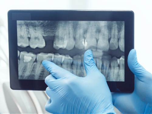
Digital X-Rays and Sensors
Digital x-rays are one of the most valuable tools we use to understand what's happening inside your mouth. Unlike traditional film x-rays, digital sensors capture detailed images of your teeth, roots, and surrounding bone in seconds. They also emit significantly less radiation, making them a healthier choice for you and your family.
Dr. Maddahi uses these images to detect cavities between teeth, evaluate the fit of crowns and fillings, and monitor bone levels in patients with gum disease. Because the images appear on screen right away, he can walk you through exactly what he sees and explain his recommendations in a way that makes sense to you. When you understand your oral health, you can make more confident decisions about your care.
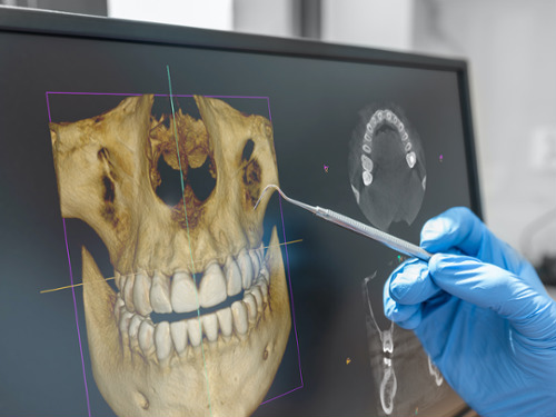
CBCT Cone Beam Technology
Planning a successful dental implant or evaluating jaw joint concerns requires a clear, complete picture of your oral anatomy. CBCT (cone beam computed tomography) technology gives Dr. Maddahi exactly that: a precise 3D image of your teeth, jawbone, nerves, and sinuses all at once. This level of detail improves treatment planning, so there are fewer surprises and better outcomes for you.
The scan itself is quick and comfortable. You simply sit or stand still for less than a minute while the scanner captures a full 3D view of the area being evaluated. There are no uncomfortable impressions or lengthy procedures involved.
Beyond implant planning, CBCT imaging supports a range of diagnostic needs. Dr. Maddahi uses these detailed scans to assess the jaw joints when temporomandibular joint (TMJ) concerns are present and to evaluate the airway for patients being assessed for sleep apnea. Having this technology in-house means you receive more thorough, informed care right here in our office.
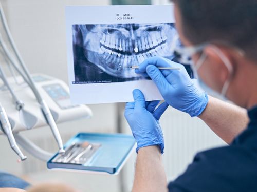
Panoramic X-Rays
A panoramic x-ray captures your entire mouth in a single, detailed image, including all your teeth, both jaws, and the jaw joints. Unlike smaller x-rays that show just a few teeth at a time, this wider view gives Dr. Maddahi a complete picture of your oral health in one easy step.
The process is quick and comfortable. You simply sit still for a few seconds while the machine gently moves around your head.
This broader view is especially useful for checking the position of wisdom teeth, monitoring how a child's adult teeth are developing, and identifying bone changes, cysts, or other concerns that smaller x-rays may not reveal. It's one of the many ways our team works to give you thorough, proactive care from the very first visit.
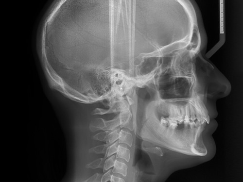
Cephalometric (Ceph) X-Rays
For children using the HealthyStart® system and patients considering Invisalign, the relationship between the teeth, jaws, and skull plays an important role in planning effective treatment. A cephalometric (ceph) x-ray is a side-view image of the head that gives Dr. Maddahi a clear picture of how these structures align and interact.
Dr. Maddahi uses ceph x-rays to measure jaw growth, assess bite concerns such as a deep overbite, and evaluate airway space. These detailed measurements guide treatment planning and let him track how a child's jaw develops over time, helping ensure each patient receives care tailored to their unique anatomy.
Rotary Endodontics
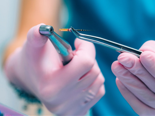
When infection or deep decay affects the soft tissue inside a tooth called the pulp, a root canal removes the damaged material and seals the tooth to protect it. Rotary endodontics is a modern approach that uses an electric handpiece and flexible nickel-titanium files to thoroughly clean the canals inside each root.
Because these files are designed to follow the natural curve of the root, they work more efficiently than traditional hand instruments. For you, this often means a shorter appointment, less time with your mouth open, and a more comfortable experience overall, which matters most when you are already dealing with a painful tooth.
Intraoral Cameras
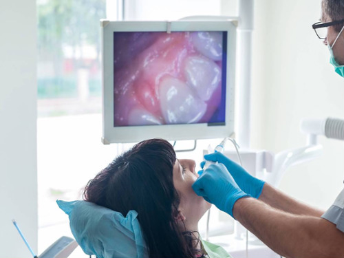
Most people have never seen the back of their own molars. An intraoral camera changes that. This small, pen-shaped device captures detailed photos of your teeth and displays them on a monitor, giving you a clear, close-up view of your own smile.
Dr. Maddahi uses these images to walk you through exactly what he sees, including cracks, worn fillings, early decay, and gum inflammation. By comparing your images to healthy teeth, he helps you understand what needs attention now and what can simply be monitored over time. When you can see the issue for yourself, treatment decisions feel less overwhelming and more informed.
Intraoral cameras also support more accurate diagnoses and help us track changes in your oral health from visit to visit. It is one more way we invest in technology that keeps you informed, comfortable, and confident in your care.
TVs in Treatment Rooms
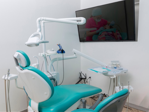
Our treatment rooms have TVs, so you can watch something to take your mind off the work happening around you. This can be especially helpful during longer appointments, such as implant placement, crown preparation, or deep cleanings for gum disease. We have found that for anxious patients, having something else to focus on can help ease their tension.